G protein
GPCR pathway is likely.
AP Biology · Unit 4 Phase 2 Deep Dive
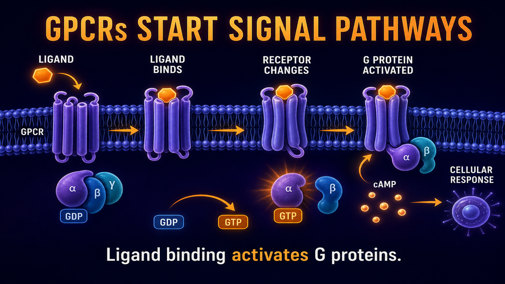
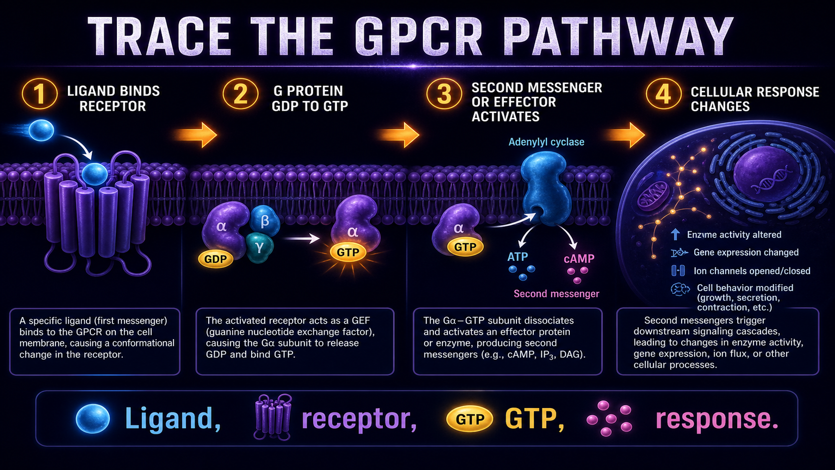
G protein-coupled receptors, or GPCRs, are membrane receptors that activate intracellular G proteins after ligand binding. When a ligand binds, the receptor changes shape, the G protein switches from GDP to GTP, and downstream effectors can produce second messengers such as cAMP. In AP Biology Unit 4, the key skill is tracing the pathway and predicting what happens when one step is blocked.
The core Unit 4 pages explain cell communication, ligands, receptors, and signal transduction. This Phase 2 deep dive focuses on one important receptor type: the G protein-coupled receptor. GPCRs are useful because they connect receptor shape change, G protein activation, second messengers, signal amplification, and response prediction.
G Protein-Coupled Receptors
G proteins relay signals inside the cell.
Also review second messengers and signal amplification as you trace full GPCR pathways.
G protein-coupled receptors are membrane receptors that activate G proteins after a ligand binds. Ligand binding changes the receptor shape, which causes the G protein to exchange GDP for GTP and activate downstream effectors. AP Biology tests GPCRs by asking students to trace the signaling pathway and predict what happens when ligand binding, G protein activation, or second messenger production is disrupted.
GPCRs use G proteins to relay signals inside the cell.
Toggle each step in order to build the GPCR pathway:
No pathway response.
Response: No response
G protein-coupled receptors are membrane proteins that detect extracellular signals and activate G proteins inside the cell. They do not usually carry the signal all the way to the final response by themselves. Instead, they start a transduction pathway that uses G proteins, effectors, and sometimes second messengers.
Connect this idea to cell communication and cell signaling pathways when you build full pathway maps on FRQs.
A GPCR is a receptor that activates a G protein after ligand binding.
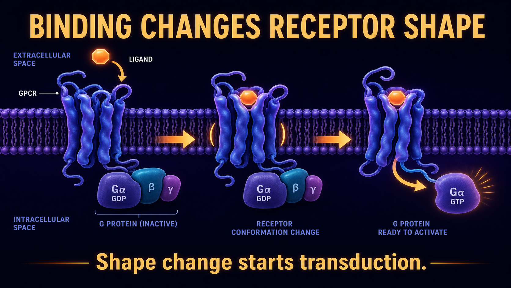
The pathway begins when a ligand binds to the outside of the GPCR. This binding changes the receptor's shape. The shape change matters because it allows the receptor to interact with and activate the G protein on the inside of the cell.
Review ligands and receptors when you need to explain why only target cells with matching receptors respond.
G proteins act like molecular switches. In the inactive state, a G protein is bound to GDP. When the activated GPCR interacts with it, the G protein releases GDP and binds GTP, switching into an active state.
G proteins are off with GDP and on with GTP.
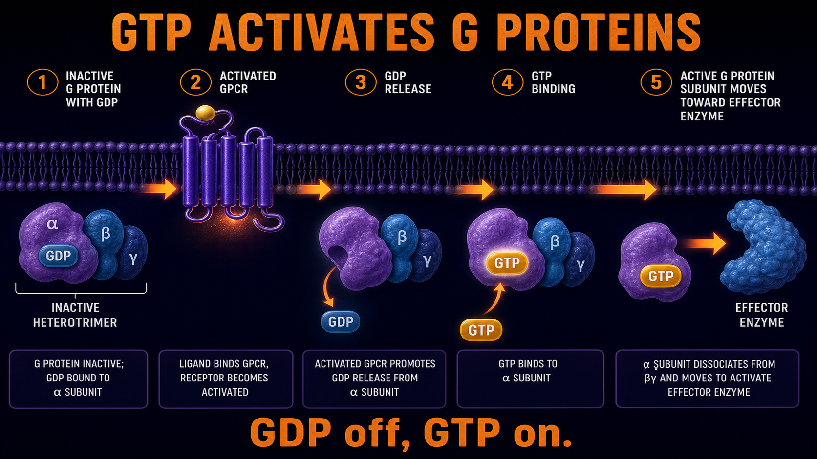
GDP and GTP help control whether the G protein is active. GDP-bound G protein is inactive, while GTP-bound G protein is active. AP Biology questions may ask what happens if the G protein cannot bind GTP: the downstream pathway will likely fail to activate.
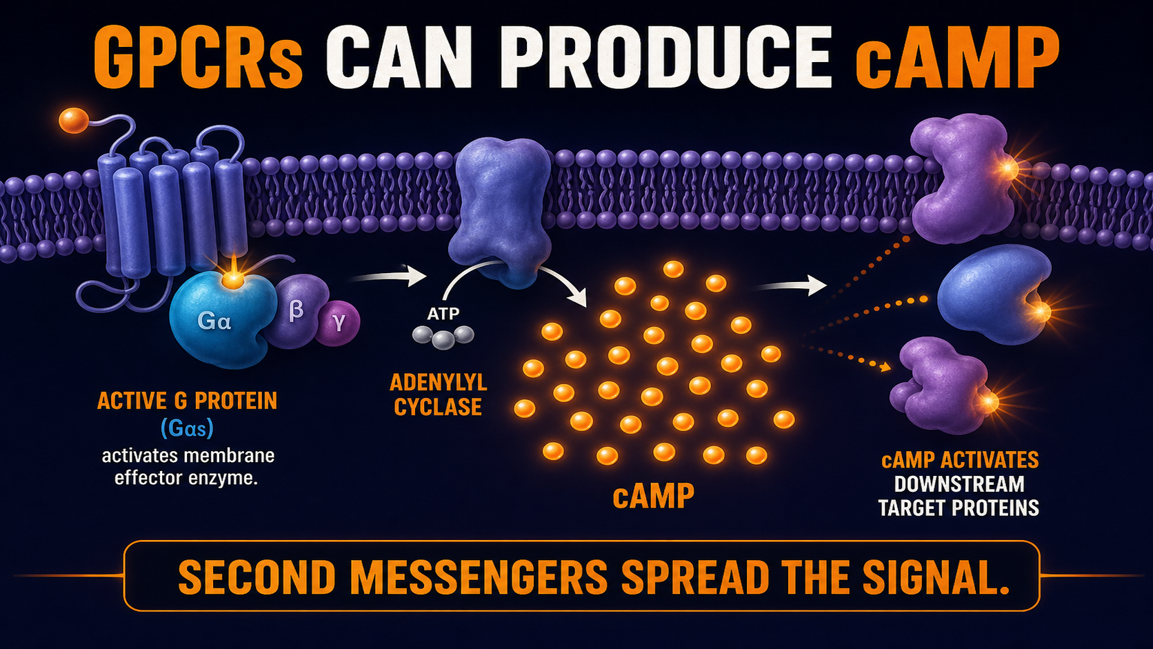
An active G protein can activate an effector enzyme in the membrane. That enzyme may produce second messengers such as cAMP. Second messengers spread the signal inside the cell and can activate many downstream proteins.
See the second messengers guide for cAMP, calcium, and other relay molecules.
One common GPCR outcome is the cAMP signaling pathway, where active G proteins stimulate adenylyl cyclase to produce cAMP.
GPCR pathways can amplify signals because one receptor event can activate G proteins, effectors, second messengers, and downstream proteins. cAMP can be produced in many copies and activate multiple targets. This helps explain how a small extracellular signal can create a large cellular response.
Read signal amplification for how each step multiplies the message.
GPCR signaling must turn off so the cell does not respond forever. G proteins can hydrolyze GTP back to GDP, returning to an inactive state. Second messengers can also be broken down, and receptors can become less responsive after prolonged signaling.
GPCRs are one major receptor type, but AP Biology may also mention receptor tyrosine kinases, ion channel receptors, or intracellular receptors. The key is to identify how the receptor changes the inside of the cell. GPCRs use G proteins; receptor tyrosine kinases often dimerize and phosphorylate; ion channels open or close; intracellular receptors regulate gene expression. Unlike GPCRs, intracellular receptors bind ligands inside the cell and often regulate transcription. Unlike a GPCR, an ion channel receptor changes cell activity by opening or closing a pore for ions instead of activating a G protein.
Compare with receptor tyrosine kinases, which usually dimerize and phosphorylate tyrosine residues instead of activating a G protein.
| Receptor type | Main mechanism | AP clue |
|---|---|---|
| GPCR | Activates G protein | GDP/GTP, cAMP |
| Tyrosine kinase receptor | Dimerizes and phosphorylates | Kinase, phosphate |
| Ion channel receptor | Opens or closes channel | Ions move |
| Intracellular receptor | Binds ligand inside cell | Steroid, gene expression |
GPCR pathway is likely.
G protein activation is being tested.
Second messenger signaling may follow GPCR activation.
Ligand binding has started transduction.
Active G protein may trigger downstream signaling.
The pathway likely stops before downstream response.
Name the signal and receptor type.
Connect binding to transduction.
Show the molecular switch turns on.
Finish with a clear outcome.
When ___ binds the GPCR, the receptor ___. The G protein exchanges ___ for ___. This activates ___, causing ___.
Fix: Many ligands bind outside the cell and activate internal pathways through receptors.
Fix: GDP is inactive; GTP is active for G proteins.
Fix: GPCRs activate G proteins; RTKs usually phosphorylate after dimerization.
Fix: Many GPCR pathways use cAMP or other second messengers.
Fix: GTP hydrolysis and messenger breakdown help stop the response.
Fix: If downstream steps are blocked, the final response may decrease or disappear.
Answer all eight questions. Choices shuffle on reload—trace the pathway, not the letter.
More drills: Unit 4 practice questions or the Unit 4 FRQ guide.
Open each card, draft your response, then reveal the rubric and sample.
A hormone binds to a GPCR on a target cell. The receptor activates a G protein, which activates an effector enzyme that increases cAMP levels.
When the hormone binds the GPCR, reception occurs because the ligand matches the receptor. The receptor changes shape and interacts with the G protein, causing GDP to be released and GTP to bind, which activates the G protein. The active G protein can then activate an effector enzyme that increases cAMP. If the G protein cannot bind GTP, it may stay inactive, the effector may not activate, cAMP may not rise, and the final cellular response may be weak or absent.
Status: Draft your answer first—then open the rubric or sample.
A mutation causes the effector enzyme in a GPCR pathway to remain active after the ligand is gone.
If the effector stays active after the ligand is removed, it may keep producing second messengers such as cAMP even when the signal should stop. High cAMP can continue activating downstream proteins, so the cellular response may last too long or become too strong. Pathways must turn off so cells do not respond forever. G proteins can hydrolyze GTP back to GDP, and second messengers can be broken down, returning the system toward its resting state.
Status: Draft your answer first—then open the rubric or sample.
G protein-coupled receptors are membrane receptors that activate G proteins after a ligand binds. The receptor changes shape and helps the G protein switch from GDP to GTP. This starts an intracellular signal transduction pathway.
A G protein acts like a molecular switch in a signaling pathway. It is usually inactive when bound to GDP and active when bound to GTP. Once active, it can trigger effectors that continue the pathway.
Ligand binding changes the shape of the GPCR. That shape change allows the receptor to activate a nearby G protein. The signal is then passed inside the cell.
GDP and GTP control whether the G protein is off or on. GDP-bound G protein is inactive, while GTP-bound G protein is active. A GPCR helps the G protein exchange GDP for GTP.
Active G proteins can activate effector enzymes that produce second messengers. cAMP is a common example. These second messengers spread the signal inside the cell.
One activated receptor can lead to many downstream signaling molecules. Effector enzymes can produce many second messenger molecules, and those messengers can activate many targets. This makes the final response larger than the original signal.
G proteins can hydrolyze GTP back to GDP, returning to an inactive state. Second messengers can also be broken down. These shutoff steps prevent the pathway from staying active too long.
If the G protein cannot bind GTP, it may not become active. The downstream effector may not activate, and second messenger production may decrease. The final cellular response may be weak or absent.
GPCRs activate G proteins, while receptor tyrosine kinases usually dimerize and phosphorylate proteins. Both can start signal transduction pathways. The mechanism is the main difference AP Biology students should track.
Trace the pathway in order: ligand binding, receptor shape change, G protein activation, effector or second messenger activation, and final response. Include GDP-to-GTP exchange if the prompt mentions G proteins. Then predict what changes if one step is blocked.