Thinking proteins are only enzymes
Fix: Many enzymes are proteins, but proteins also transport, signal, support, move, defend, and act as receptors.
AP Biology · Unit 1 · Chemistry of Life
Learn how amino acid chains fold into enzymes, transport proteins, receptors, and structural molecules.
AP Biology proteins questions usually test whether you can connect amino acid sequence to protein shape and protein function. Proteins are built from amino acids, joined by peptide bonds, folded into specific shapes, and used for enzymes, transport, structure, signaling, receptors, and movement. This page explains protein structure, folding, denaturation, and AP-style clues.
Unit 1 progress
AP Biology · Unit 1
Part of Unit 1: Chemistry of Life · Page 8 of 11
Pair with lipids (previous) and nucleic acids (next).
Proteins are biological macromolecules made from amino acid monomers. Amino acids join by peptide bonds to form polypeptides, and polypeptides fold into specific shapes. In AP Biology, proteins are important because their shape allows them to function as enzymes, transport proteins, receptors, structural molecules, signaling molecules, and more.
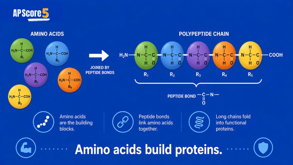
AP Biology proteins are amino acid-based macromolecules that fold into specific shapes and perform many cell functions. Proteins can act as enzymes, transport proteins, receptors, structural molecules, signaling molecules, and movement-related molecules. The most important AP Biology idea is that protein function depends on protein shape. Amino acid sequence affects folding, folding affects shape, and shape affects function. If a protein's shape changes because of heat, pH, salt concentration, or mutation, its function can change too.
Proteins AP Biology content sits after lipids and carbohydrates in the Unit 1 macromolecule sequence because proteins are true polymers with a clear monomer-polymer story. Unlike lipids, which often break the repeating-monomer pattern, proteins follow the classic build logic you practiced on monomers and polymers and dehydration synthesis and hydrolysis. Place this page in the broader macromolecules comparison when you need to contrast all four classes, or return to the Unit 1 Chemistry of Life hub for the full learning path.
Water chemistry still matters here. Polar and nonpolar R groups interact differently with aqueous cytosol, which drives folding patterns you first met on the water properties page. Nitrogen in amino groups and sulfur in some side chains link back to elements of life. Strong Unit 1 protein study connects those earlier topics to the four levels of structure, enzyme active sites, and membrane transport jobs you will see again in Unit 2, Unit 3, and Unit 6 gene expression.
Proteins are one of the most important macromolecule groups in AP Biology because they perform so many cell jobs. Proteins can act as enzymes, transport proteins, receptors, structural molecules, signaling molecules, and movement-related molecules. If a cell is doing work, there is a good chance proteins are involved.
In AP Biology, proteins matter because their amino acid sequence affects folding, and folding affects function.
This structure-function chain is the most important idea on this page. Proteins are built from amino acids. The order of amino acids affects how a protein folds. The folded shape affects what the protein can do. If the shape changes, the function can change or be lost.
That is why AP Biology questions often ask about protein folding, denaturation, enzymes, active sites, membrane proteins, or mutations that change amino acid sequence. The details may look different, but the reasoning is often the same: structure affects function. AP questions expect you to trace amino acid sequence → folding → shape → function → cell process in both multiple-choice stems and free-response answers.
Proteins also connect Unit 1 to later units without turning this page into a full protein synthesis lesson. Enzymes in cellular respiration, receptors on membranes, and hemoglobin in blood all assume you understand how sequence and folding create functional shape. DNA and RNA store and transmit the information cells use to build proteins—a connection you will deepen on the nucleic acids page and in Unit 6—but the Unit 1 focus stays on structure and function, not ribosomes and codons.
Compare proteins with other macromolecules when a question lists all four classes. Carbohydrates mention glucose and polysaccharides; lipids mention hydrophobic tails and bilayers; nucleic acids mention nucleotides and bases. When a stem highlights peptide bonds, active sites, or denaturation, proteins should move to the top of your answer list before you read all four choices.
Proteins contain carbon, hydrogen, oxygen, nitrogen, and sometimes sulfur. Proteins are built from amino acid monomers. Amino acids form polypeptides through peptide bonds, and one or more polypeptides fold into a functional protein. Unlike lipids, proteins are true polymers where repeating monomers link in a defined sequence—and that sequence matters for everything downstream.
Nitrogen appears in amino groups on every amino acid, which is why protein questions often contrast C, H, O-rich carbohydrates with nitrogen-containing polypeptides. Sulfur appears in some R groups and enables disulfide bonds that stabilize tertiary structure in extracellular and membrane-facing proteins. Those element clues narrow answer choices quickly when a multiple-choice item asks which macromolecule class contains nitrogen or sulfur.
| Feature | Protein Pattern | AP Biology Meaning |
|---|---|---|
| Main elements | C, H, O, N, sometimes S | Nitrogen appears in amino groups; sulfur can stabilize some proteins |
| Monomer | Amino acid | Building block of proteins |
| Polymer | Polypeptide / protein | Chain that folds into a functional molecule |
| Bond | Peptide bond | Links amino acids during dehydration synthesis |
| Key idea | Shape determines function | Folding affects protein job in the cell |
A functional protein may contain one polypeptide or several subunits held in quaternary structure. Hemoglobin is a classic AP example: four polypeptide chains cooperate to bind and release oxygen. Collagen forms structural fibers in connective tissue. Amylase in saliva is a single-chain enzyme that breaks starch. The common thread is amino acid sequence encoded in the chain, folded into a shape that fits a biological job.
When you compare proteins to lipids on the lipids study guide, remember that lipids are grouped mainly by hydrophobic behavior while proteins are grouped by amino acid polymers with sequence-dependent folding. Both are macromolecules, but the exam tests different structure-function patterns for each class.
Amino acids are the monomers of proteins. Each amino acid has the same basic framework: a central carbon, an amino group, a carboxyl group, a hydrogen atom, and an R group. The R group is the variable part. It gives each amino acid its chemical personality and strongly influences how the finished protein folds.
This matters because R groups affect folding. Some R groups are nonpolar and tend to avoid water in the interior of a folded protein. Some are polar or charged and interact well with the aqueous environment on the protein surface. Some contain sulfur and can form disulfide bridges that lock tertiary structure in place. These interactions help determine the final 3D shape of the protein and therefore its function.
For AP Biology, you do not usually need to memorize every amino acid structure, but you should understand why amino acid properties affect protein folding and function. When a mutation replaces one amino acid with another, the new R group chemistry can shift folding enough to change an enzyme active site or a receptor binding pocket.
| Amino Acid Part | What It Means | Why AP Biology Cares |
|---|---|---|
| Central carbon | Core atom of amino acid | Holds the amino acid parts together |
| Amino group | Contains nitrogen | Helps identify amino acids and peptide bond formation |
| Carboxyl group | Acidic functional group | Participates in peptide bond formation |
| R group | Variable side chain | Gives each amino acid different properties |
| Hydrogen | Attached to central carbon | Part of general amino acid structure |
Think of twenty common amino acids as twenty different side-chain personalities arranged in a specific order along a polypeptide backbone. That order is primary structure. The R groups then push and pull the chain into helices, sheets, and loops until a stable 3D shape emerges. AP free-response questions love that logic because it rewards understanding over memorization of structural diagrams.
Amino acids join by peptide bonds. Peptide bonds form through dehydration synthesis: the carboxyl group of one amino acid reacts with the amino group of the next, releasing water and creating a covalent link between the two residues. Repeat that reaction dozens or hundreds of times and you build a polypeptide—a true polymer chain with directionality from N-terminus to C-terminus.
Simple flow to remember: amino acid + amino acid → dipeptide + water. Scale up: amino acids → polypeptide → folded protein → function. Hydrolysis reverses the process by adding water to break peptide bonds, splitting a polypeptide into amino acids. Digestive enzymes use hydrolysis to break dietary proteins into absorbable units; cells also use controlled hydrolysis to recycle damaged proteins.
This links directly to the dehydration synthesis and hydrolysis page. The same build-and-break logic applies to glycosidic bonds in carbohydrates and phosphodiester bonds in nucleic acids—only the monomers differ. Peptide bonds are the protein-specific linkage AP Biology expects you to name on MCQs and FRQs.
Do not confuse peptide bonds with glycosidic bonds (sugars) or phosphodiester bonds (nucleic acids). If a question asks which bond joins amino acids, the answer is always peptide bond. If a question describes two monosaccharides joining, that is glycosidic chemistry on the carbohydrates page, not protein chemistry here.
Protein structure has four levels, and AP Biology students should understand the logic behind each one. Primary structure is the amino acid sequence—the starting point written in order along the chain. If the sequence changes, folding may change, and function may follow.
Secondary structure refers to local folding patterns such as alpha helices and beta sheets stabilized mainly by hydrogen bonds between backbone atoms. Tertiary structure is the overall 3D shape of one polypeptide, driven by R group interactions including hydrophobic clustering, ionic attractions, hydrogen bonds, and disulfide bridges. Quaternary structure occurs when multiple polypeptide subunits assemble into one functional protein complex.
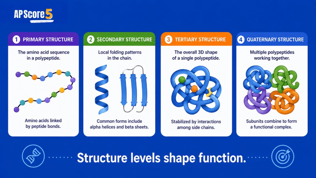
| Level | What It Means | What Stabilizes It | AP Biology Test Clue |
|---|---|---|---|
| Primary | Amino acid sequence | Peptide bonds | Sequence, order of amino acids, mutation |
| Secondary | Local folding patterns | Hydrogen bonds | Alpha helix, beta sheet, local folding |
| Tertiary | Overall 3D shape of one polypeptide | R group interactions, hydrophobic interactions, ionic bonds, disulfide bonds | Shape, active site, folding |
| Quaternary | Multiple polypeptide subunits | Interactions between subunits | Multi-subunit protein, hemoglobin-like example |
The AP Biology focus is not memorizing labels alone. The focus is explaining how changes in structure can affect function. If a mutation changes an amino acid, that can change folding. If folding changes, the active site or binding region may change. If shape changes, function may decrease or disappear. That causal chain appears in denaturation scenarios, enzyme inhibition contexts, and genetic mutation FRQs across the course.
Secondary structure is often the first folding step after the ribosome releases a new chain. Tertiary structure packs those local motifs into a compact globular or fibrous final form. Quaternary structure adds cooperation between chains—oxygen binding in hemoglobin depends on all four subunits working together. Use the structure explorer below to review each level interactively before the flashcards and MCQs.
Protein folding creates the functional shape a cell needs. Folding depends on amino acid sequence and R group interactions with each other and with the surrounding aqueous environment. Nonpolar R groups often bury themselves in the protein interior; polar and charged R groups often face outward toward water. Those patterns mirror the hydrophobic effect you studied with lipids and water.
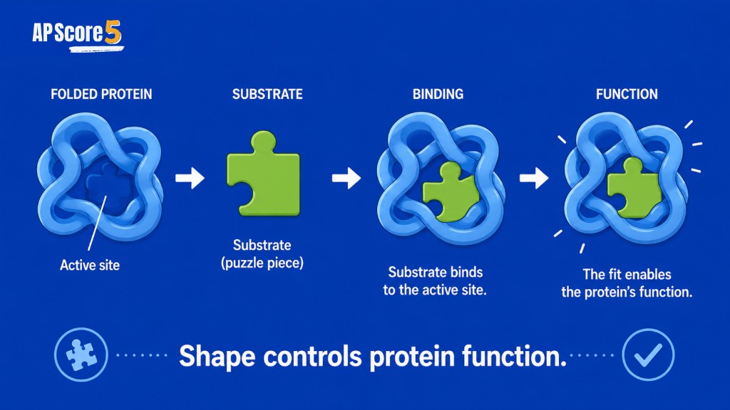
Protein shape affects binding. Enzymes need specific active sites shaped to fit substrates. Receptors need binding pockets matched to signal molecules (ligands). Transport proteins need channels or carrier conformations that move only certain ions or molecules. Antibodies need variable regions that recognize specific antigens. In every case, the 3D geometry—not just the amino acid list—determines whether the interaction succeeds.
Folding is sensitive to environment. pH changes can alter charged R groups and disrupt ionic bonds holding tertiary structure. Temperature increases can add kinetic energy that breaks weak interactions. Salt concentration can shield charges and reshape ionic networks. These environmental effects set up the denaturation section next and explain why cells tightly regulate conditions in cytosol, blood, and organelle compartments.
Structure-function chain to write on FRQs: amino acid sequence → R group interactions → folding → 3D shape → function. If any link breaks, the protein may still exist as a chain of amino acids but fail at its job.
Denaturation is a change in protein shape. Heat, pH shifts, salt concentration changes, or harsh chemicals can disrupt the interactions that maintain secondary, tertiary, and quaternary structure. Denaturation may reduce or destroy function even when the primary amino acid sequence—and peptide bonds—remain intact.
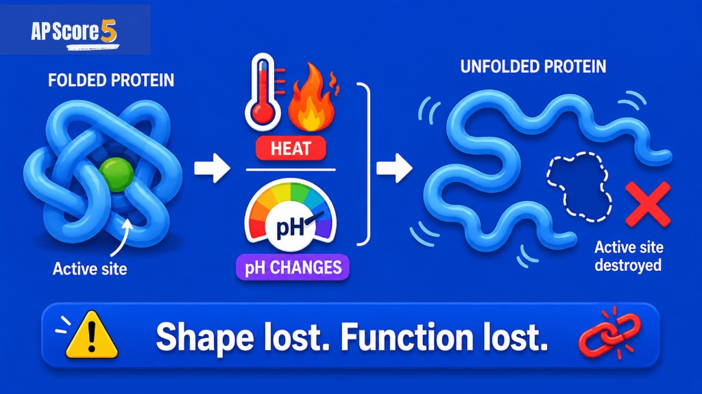
Enzymes are the classic AP example. Boiling an enzyme in a lab often permanently reduces activity because the active site loses its complementary shape. Cooking egg white denatures albumin proteins, turning clear liquid into solid white—that is visible evidence of shape change without breaking every peptide bond. Cells avoid uncontrolled denaturation with chaperone proteins and stable pH buffers, but extreme conditions still appear in exam scenarios.
| Cause | What It Can Disrupt | AP Result |
|---|---|---|
| High temperature | Weak interactions and folding | Protein shape changes; enzyme rate may drop |
| pH change | Charge interactions on R groups | Active site may change shape |
| Salt concentration | Ionic interactions | Folding may change |
| Chemical environment | R group interactions | Function may decrease |
Distinguish denaturation from hydrolysis. Denaturation alters shape while the polypeptide chain largely stays intact. Hydrolysis breaks peptide bonds and produces amino acids or shorter fragments—a chemical digestion process, not merely unfolding. Both can stop a protein from working, but the mechanisms differ and AP questions may ask you to choose between them.
Proteins are not one-job molecules. Their diversity comes from amino acid sequence and folding. A protein's shape allows it to do a specific job. That is why proteins can be enzymes, receptors, transport proteins, structural proteins, signaling molecules, motor proteins, defensive antibodies, and storage proteins—all built from the same monomer set arranged differently.
| Protein Function | Example | Why It Matters |
|---|---|---|
| Enzymes | Amylase, catalase | Speed up chemical reactions |
| Transport | Membrane channels, hemoglobin | Move substances within or between compartments |
| Structure | Collagen, cytoskeleton proteins | Provide support and cell shape |
| Signaling | Hormones or signal proteins | Coordinate cell and organism responses |
| Receptors | Membrane receptors | Detect chemical signals |
| Movement | Motor proteins | Support movement in cells and organisms |
| Defense | Antibodies | Help identify pathogens |
| Storage | Some storage proteins | Store molecules or ions |
When an AP prompt lists a molecule that speeds a reaction, think enzyme and therefore protein. When it describes oxygen delivery in blood, think hemoglobin and quaternary structure. When it mentions cell wall-like support in animals, think collagen fibers rather than cellulose (a carbohydrate in plants). Matching function words to protein examples helps you eliminate carbohydrate and lipid distractors quickly.
Proteins also appear as glycoproteins on cell surfaces, helping recognition and adhesion—topics that bridge to Unit 2 membrane biology. You do not need full signaling cascades here; recognize that receptor shape determines which ligand fits, just as enzyme shape determines which substrate fits.
Enzymes are one of the most important protein examples in AP Biology. An enzyme speeds up a chemical reaction by lowering activation energy. Its shape matters because the active site must fit or interact with the substrate—the reactant the enzyme acts on. Induced fit models describe how the active site and substrate adjust slightly upon binding, but the core idea remains: complementary shape enables catalysis.
Denaturation connects directly to enzymes. Heat or pH changes can alter protein shape. If an enzyme's active site changes, the enzyme may no longer bind the substrate effectively, and the reaction rate may decrease even though substrate concentration stays high. That pattern foreshadows enzyme kinetics and regulation in Unit 3 cellular energetics, but the Unit 1 takeaway is structure-function at the active site.
Not every catalyst in biology is a protein—some RNA molecules have enzymatic activity—but AP Biology MCQs and FRQs most often test protein enzymes such as amylase, pepsin, DNA polymerase, and catalase. When a stem names an enzyme, default to protein unless the question explicitly describes ribozyme RNA.
Proteins are essential in membranes. While phospholipids form the bilayer fabric you studied on the lipids page, many membrane jobs depend on embedded or surface proteins. Channel proteins provide passageways for ions and small polar molecules. Carrier proteins change shape to transport substances that cannot diffuse freely through the hydrophobic interior. Receptor proteins bind signaling molecules and initiate cellular responses.
Protein shape helps determine what can bind or pass through. A sodium channel selects Na⁺ based on size and charge at its pore. A glucose carrier opens to one side of the membrane, binds glucose, then shifts conformation to release it on the other side. A receptor for a peptide hormone exposes an extracellular binding domain matched to that hormone's shape. Each example is structure-function reasoning applied to the membrane context you will expand in Unit 2 cell structure and function.
Many membrane proteins have hydrophobic regions that anchor in the lipid bilayer and hydrophilic regions that contact aqueous cytosol or extracellular fluid—an amphipathic arrangement parallel to phospholipids, but with amino acid R groups instead of fatty acid tails. Integral and peripheral membrane proteins differ in attachment strength, but AP Unit 1 usually stops at the concept that membranes are lipid-plus-protein systems, not lipid alone.
Structure-function reasoning is the scoring language of AP Biology protein questions. The exam wants you to connect sequence, R group chemistry, folding level, and biological outcome—not merely define vocabulary in isolation.
| Pattern | Protein Feature | Function | AP Test Clue |
|---|---|---|---|
| Amino acid sequence | Primary structure | Determines folding potential | Mutation, sequence change |
| R group interactions | Side chain chemistry | Drives folding | Polar, nonpolar, charged, sulfur |
| Active site shape | Tertiary structure | Enzyme binding and catalysis | Substrate, reaction rate |
| Membrane channel shape | Folded protein | Selective transport | Selective passage, ion flow |
| Receptor shape | Binding site | Signal detection | Ligand, signal molecule |
| Denaturation | Shape disruption | Function loss | Heat, pH, salt |
| Disulfide bond | Sulfur interaction | Stabilizes shape | Cysteine, sulfur, tertiary structure |
Practice translating structures into verbs: catalyze, bind, transport, signal, support, move, defend, store. If your FRQ answer uses only nouns (enzyme, receptor, hemoglobin), add a verb that states what the cell accomplishes with that structure. That habit aligns your writing with official scoring guidelines and helps you earn the third point on many three-point prompts.
Connect patterns across units without leaving Unit 1 focus. A mutation changing one amino acid is a nucleic acid story later, but the immediate AP payoff is folding and function here. An enzyme denatured by fever is a protein shape story before it is a physiology story. Keep the chain visible in every answer.
Protein questions rarely announce themselves with the word protein. Instead they describe amino acids, peptide bonds, folding levels, enzymes, active sites, denaturation, receptors, or transport across membranes. Train yourself to recognize vocabulary clusters and map each cluster to structure-function logic before you read answer choices.
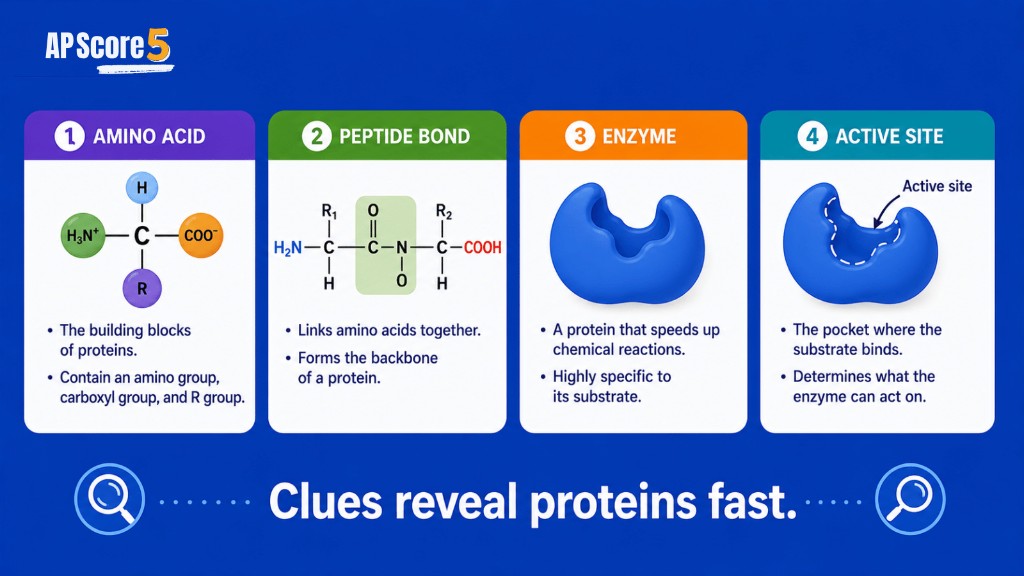
Distinguish protein structure questions from protein synthesis questions. If a stem mentions ribosomes, codons, transcription, or translation, you may be in Unit 6 territory. If it mentions folding, denaturation, active sites, or four structure levels, stay in Unit 1 protein chemistry. Both use the word protein, but the reasoning path differs.
Practice this checklist with the shuffled MCQs below—letter positions change each load, so you must think in concepts, not memorized answer keys. Additional drills live on practice by topic and daily AP Biology practice. The AP Biology course page links all units when you want a wider review.
Fix: Many enzymes are proteins, but proteins also transport, signal, support, move, defend, and act as receptors.
Fix: Amino acids build polypeptides, which fold into proteins.
Fix: Peptide bonds link amino acids. Glycosidic bonds link sugars.
Fix: Protein shape is central to function.
Fix: Denaturation changes shape; hydrolysis breaks bonds into smaller subunits.
Fix: R groups give amino acids different properties and affect folding.
Fix: Primary is sequence, secondary is local folding, tertiary is overall 3D shape, quaternary is multiple subunits.
Fix: Protein synthesis comes later; this Unit 1 page focuses on protein structure and function.
Open each card to reveal the answer and why the clue fits. Use these before the structure level explorer below.
Answer: Primary structure
Primary structure is the order of amino acids.
Answer: Secondary structure
Secondary structure describes local folding patterns.
Answer: Tertiary structure
Tertiary structure is the full folded shape of one polypeptide.
Answer: Quaternary structure
Quaternary structure involves more than one polypeptide chain.
Answer: Denaturation
Heat can change protein shape and reduce function.
Answer: Peptide bond
Peptide bonds link amino acids into polypeptides.
Answer: R group
R groups give amino acids different properties.
Answer: Enzyme
Enzymes are proteins that catalyze reactions.
Answer: Receptor
Receptors bind signal molecules.
Answer: Transport protein
Channels and carriers help substances cross membranes.
Tap each structure level once to open details. Explore all four to enable the finish button sooner.
Amino acid sequence · peptide bonds · starting point for folding
What it is: The linear order of amino acids in a polypeptide
Stabilized by: Peptide (covalent) bonds between amino acids
AP clue: Sequence, mutation, order of amino acids
Local folding · alpha helices · beta sheets · hydrogen bonds
What it is: Repeating local patterns such as coils and pleated sheets
Stabilized by: Hydrogen bonds between backbone atoms
AP clue: Alpha helix, beta sheet, local folding
Overall 3D shape · R group interactions · active sites
What it is: The full folded shape of one polypeptide chain
Stabilized by: Hydrophobic interactions, ionic bonds, disulfide bonds, hydrogen bonds
AP clue: Active site, folding, shape, denaturation
Multiple subunits · hemoglobin-like proteins · subunit assembly
What it is: Two or more polypeptide chains arranged as one functional protein
Stabilized by: Interactions between separate polypeptide subunits
AP clue: Multi-subunit protein, hemoglobin, four chains
0 of 4 structure levels explored · tap each card once
Follow these steps in order. You are on step 8.
Review how all four macromolecule classes compare.
Review building blocks and true polymers.
Review dehydration synthesis and hydrolysis.
Review elements of life.
Connect proteins to membranes in Unit 2.
Review cellular energetics in Unit 3 later.
Continue to nucleic acids and Unit 6 gene expression.
Continue to nucleic acids.
Every 5th card shows an ad placeholder with a short countdown. Flip the card to read the definition, then use the arrow for the next card.
Choices shuffle at display time. Tap an answer, read the explanation, then use Next question.
Want more Unit 1 drills? Try daily AP Biology practice or practice by topic.
Click a question to open the full prompt. Write your answer on paper first, then reveal the rubric and a strong sample response.
A change in amino acid sequence can alter how the polypeptide folds. If folding changes the enzyme's active site shape, the substrate may not bind properly and enzyme function may decrease.
This is the same structure-function chain tested on Unit 1 protein pages and later in transcription vs translation when mutations affect the protein product.
Sequence → folding → active site → function. Name each link in order.
Denaturation changes a protein's folded shape by disrupting interactions that maintain structure. Since protein function depends on shape, the protein may no longer bind or interact correctly.
Heat disrupts hydrogen bonds and other weak interactions holding the tertiary structure together. The active site may lose its complementary shape even though peptide bonds in the primary sequence remain intact.
Denaturation = shape change, not disappearance of the protein molecule.
Primary structure is the amino acid sequence of a protein—the order determined by peptide bonds from the N-terminus to the C-terminus. Tertiary structure is the overall 3D shape of one polypeptide, formed by interactions among R groups and the surrounding environment.
Secondary structure (alpha helices and beta sheets) sits between them as local folding patterns. Review the four levels on the macromolecules comparison when you need to contrast all macromolecule classes.
Primary = sequence. Tertiary = full 3D fold of one chain.
Transport proteins have specific shapes that allow certain substances to pass through or bind. Channel proteins form passageways with a size and charge profile matched to specific ions or molecules. Carrier proteins change shape to move a substrate across the bilayer.
Membranes are built from phospholipids, but many membrane jobs depend on proteins—connect this to Unit 2 cell structure and the lipids bilayer page for full context.
Shape → selective passage. Name channel vs carrier when the prompt allows.
Proteins are biological macromolecules made from amino acids that fold into specific shapes and perform many cell functions.
The monomers of proteins are amino acids.
Peptide bonds link amino acids together in polypeptides.
A polypeptide is a chain of amino acids that can fold into a functional protein.
Protein shape matters because the shape determines what the protein can bind, interact with, or do.
The four levels are primary, secondary, tertiary, and quaternary structure.
Primary structure is the amino acid sequence of a protein.
Tertiary structure is the overall 3D shape of one polypeptide.
Denaturation is a change in protein shape that can reduce or destroy protein function.
Many enzymes are proteins, and their shape helps them bind substrates and speed up reactions.
Many membranes contain proteins that help transport substances, receive signals, or support cell communication.
DNA contains information that can be used to make proteins, and RNA helps carry out that process in gene expression.
Clues include amino acids, peptide bonds, polypeptides, folding, active sites, enzymes, receptors, transport proteins, and denaturation.
Be ready to explain how amino acid sequence affects folding, how folding affects shape, and how shape affects protein function.
Check each skill when you can explain it without looking at the page.
0 of 17 skills ready
Nice work—you explored all four structure levels and checked off the review skills. Continue to nucleic acids for DNA and RNA information, or return to the macromolecules overview to compare all four classes.
You just finished the proteins study guide. Next, study nucleic acids for genetic information, or review the full macromolecules comparison.